Introduction
In previous articles, we’ve looked at the very basics of inflammation and then in-depth with the specifics.
This time around, we’re going to discuss the human spine, how it is constructed and what it is capable of. To do this we are going to build a model spine involving some everyday objects.
We are doing it this way to make it easy to understand so that firstly you grasp it fully and secondly you can use the information to help to teach your clients and patients about the way their spines are built and how to promote spine health.
A Difficult Task and a Clever Solution
To understand how the spine is made up we need to think about what our spines need to do during our daily lives. The spine needs to form the connection between the head, the arms and the body. It also needs to be strong enough to take the weight of all the bones above it and still be rigid enough to form a stable platform for our arms and legs to move. Finally, it also needs to move in all directions in its own right.

Evolution has provided a satisfactory solution to this by having a large number of movable joints where each pair of bones moves a slight amount, but when these are added up, they give us a great deal of flexibility while the spine maintains stability.
Bones
The main unit of the spine is the vertebral body. These are shaped rather like marshmallows, but we will compare them to bricks.
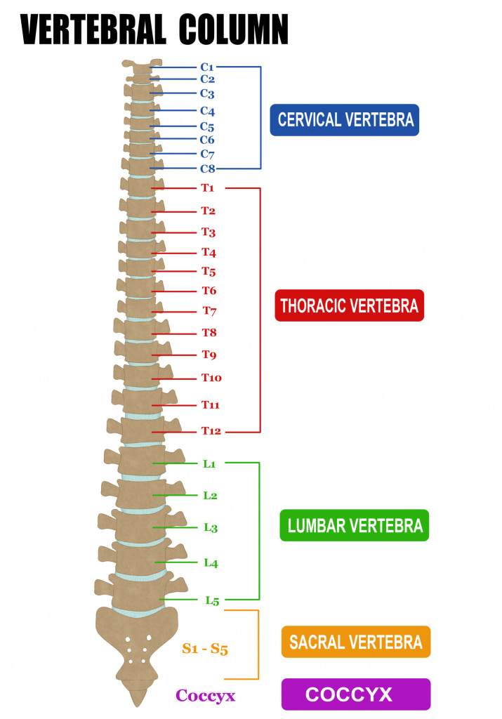
The bricks are stacked on top of each other with the head at the top of the pile and pelvis at the bottom. There are seven bricks in the cervical spine, 12 in the thoracic spine, and five in the lumbar spine. These bricks get larger as we descend since they need to carry more and more weight. They are made of cancellous bone which is very light but also very strong. Unfortunately, it is also prone to osteoporosis so it is not unusual to see a collapsed vertebra in a patient with this condition.
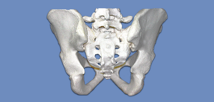
The pelvis ends in the sacrum which is a series of fused vertebra that wedge into the pelvis. The very tip of the spine is the coccyx which is the remains of our ancestors’ tails.
The Disc
The bricks are separated by intervertebral discs which we will compare to a jam doughnut. Like a jam doughnut, the disc has a hard outer shell called the annulus fibrosis and a soft jammy gel-like centre called the nucleus pulposus. These discs are flexible enough to allow the bones to rock in various directions but also strong enough to give stability.
Getting on Your Nerves
At the back of the column of bricks there is a ring of bone and through the central hole in this ring passes the spinal cord.
The ring is called the spinal canal and the spinal cord runs from the base of the brain to approximately halfway down the spine.
Controlling the Spine
We now need to think about how we can control this potentially very wobbly column. What we have is a situation where the jam doughnuts are pumped up to maximum pressure which makes them rigid and they are encased by ligaments which also limit the movements of the spine.
More and More Layers
Added onto the ring we have a series of three bony projections like lollipop sticks. Two project sideways and one goes posteriorly or backwards. The ones that go sideways are called transverse processes and these act as anchors for ligaments and muscles, and the processes that point backwards are called spinous processes.
You can feel these with your own hands when you run your hands down the back of somebody’s neck or spine. These also serve to anchor ligaments and muscles.
Curves and Movements
We now need to think about the shape of a normal spine. When a baby is born it has one curved spine making it look rather like a C shape. Once the baby starts to take an interest in things and crawl around secondary curves develop in the other parts of the spine which means that we have what is known as a cervical and lumbar lordosis and thoracic kyphosis, this gives the spine additional strength balance and manoeuvrability.
Not the Same
Different parts of the spine are specialised for different things, so for example in the neck, the bones are adapted to allow rotation and forwards and backwards movement. In the thoracic spine, some movements are a little limited by the presence of the rib cage which also joins onto the spine. In the lumbar spine rotation, it’s not desirable so we have a series of joints called facet joints which are located in such a way as to either allow or limit rotation in the spine.
Another important difference in the neck is that the top two vertebrae are very strangely shaped, they are called C1 and C2 or the atlas and the axis and they bear little resemblance to the other bones in the neck.
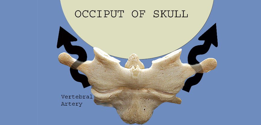
Additionally, the other cervical vertebrae have little holes in each side which allow an artery to go up through the bones to the brain. This is called the vertebral artery and for this reason, we need to be very careful about manipulating the cervical spine especially if somebody has arthritis in the spine or a history of circulatory disorders.
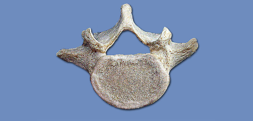
To conclude, let’s have a look at some photos that I’ve taken which bring to life the anatomy of the spine.
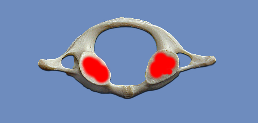
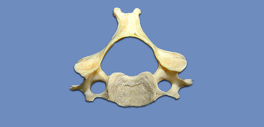
The first cervical vertebra – the skull rests on the two facets coloured red here
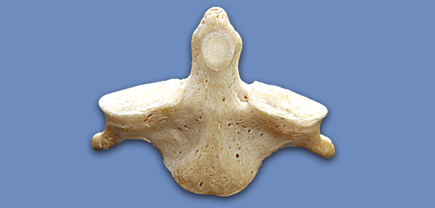
The second cervical vertebra – the peg sticking up is called the odontoid or the dens
This shows the position of the skull and the vertebral arteries and how they relate to the second cervical vertebra
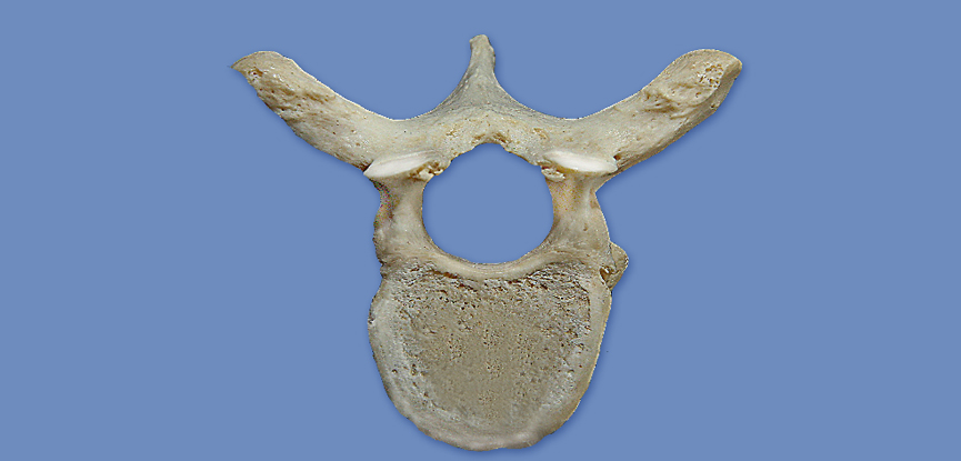
A typical cervical vertebra
A typical thoracic vertebra
A typical lumbar vertebra
The sacrum and the pelvis